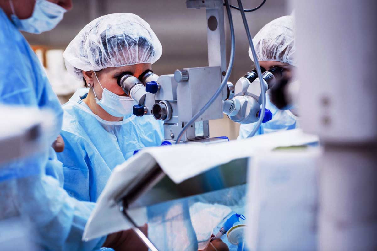
The very first non-invasive new technology, OCT-based epithelial mapping, was built for a quantitative measure of the corneal epithelial and stroma. To evaluate the change in the patient’s ocular, the eye surgeon uses OCT-based epithelial mapping to aid in the diagnosis of corneal disease.
With the help of using a map measuring 9 mm in diameter of OCT epithelial mapping, it becomes easy for the eye surgeon to map the corneal epithelium.
The process is a quick two-second scan that will help in identifying areas of thickening or thinning related to dry eye disease, keratoconus, or any previous refractive surgery. The OCT mapping distinguishes between forme fruste keratoconus, and contact lens-related warpage. This device helps the ophthalmologist to understand the anatomy of your eye so he can make the best decision for you.