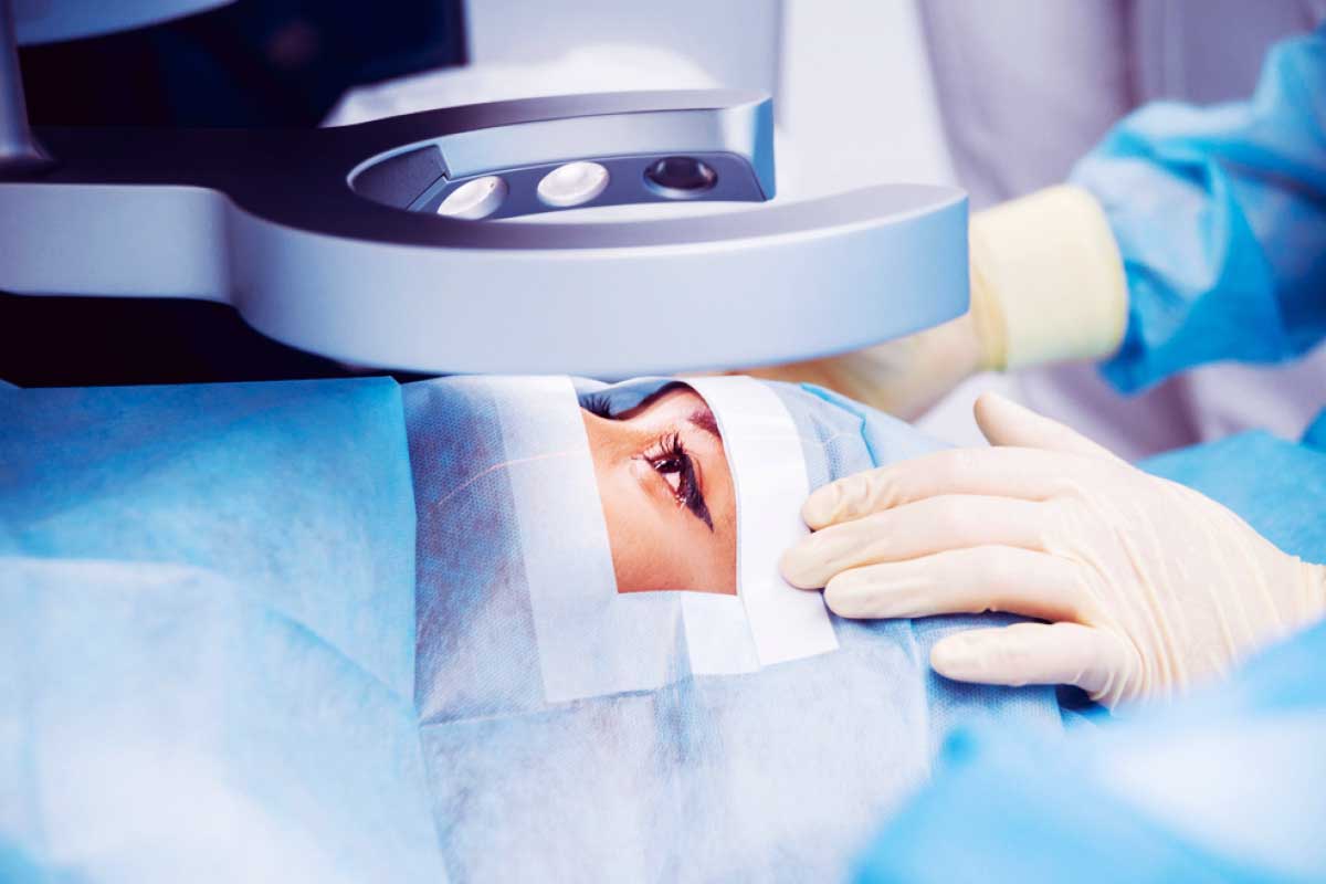
Hashmanis Group of Hospitals is known to have the authority in the field of Ophthalmology, has introduced over 15 different treatment modalities to Pakistan. Currently, Hashmanis is the only Hospital that takes the privilege to be counted among a handful of hospitals in the world to perform advanced treatments such as laser-assisted (bladeless) cataract surgery and Topography Guided LASIK. Hashmanis offers a host of specialized treatments for refractive surgeries, glaucoma, cataract, corneal, pediatric, and retinal eye problems.
We, at Hashmanis Hospital, ensure to provide routine eye exams as well as perform eye surgeries with the aid of high tech machines to promote quality vision.
Zero Compression Microkeratome
The Zero Compression Microkeratome is the new technology for laser vision correction. This device saves a patient’s corneal tissue through thinner, smoother, and more uniform flaps.
Read more
Femtosecond Lasers
The new and advanced bladeless device; Femtosecond lasers provide patients with the highest standard of precision and reliability in the creation of a corneal flap.
Read more
Excimer Lasers
Excimer lasers are the only machine that uses ultraviolet rays to reshape the cornea and also correct the refractive error of the eye at the same time.
Read more
Topography
Topography procedure is a non-invasive and painless procedure used for corneal imaging techniques. This procedure helps in determining the quality of vision, assist in LASIK surgery, and the fitting of contact lenses.
Read more
iTrace™ Aberrometer
The 5-in-1 system, the iTraceiTrace™ Aberrometer, provides a better and accurate analysis of the eye, making it easy for the ophthalmologist to interpret reports and simple to integrate.
Read more
OCT-Based Epithelial Mapping
The OCT Epithelial Mapping provides a quantitative measure of the corneal epithelial and stroma. The eye surgeon evaluates the diagnosis of corneal disease in the patient’s ocular surface.
Read more
Corneal Visualization Scheimpflug Technology (CorVis ST)
The Corneal Visualization Scheimpflug Technology (CorVis ST) helps to investigate the corneal biomechanical properties in patients with dry eye.
Read more
Dry Eye Analyzer
The quickest and safest way to examine the cause of irritability within the patient’s eye is through the Dry Eye Analyzer. The analyzer examines the cornea by combining infrared imaging, interference light measurement, and magnification technology.
Read more
A-Scan and B-Scan
Both the ultrasound scanners evaluate the posterior segment and orbital pathology, especially when the patients visual gets cloudy, and the ophthalmologist has an issue with a direct view for further diagnosing.
Read more
Verion Digital Marker
The Verion digital marker of Alcon helps in cataract surgeries. With its accurate precision, consistency, and control in cataract refractive surgery, it allows the eye surgeon to create a customized procedure according to each patient’s health by performing key diagnostic measurements and capturing a high-resolution image of the eye in a single step!
Read more
Yag Laser
YAG laser treatment is safe to use for treating medication conditions within the anterior segment of the eye. Surgeries such as glaucoma and cataract use YAG laser in their procedures.
Read more
iTrace
The 5-in-1 system, the iTraceiTrace™ Aberrometer, provides a better and accurate analysis of the eye, making it easy for the ophthalmologist to interpret reports and simple to integrate.
Read more
IOL Master
The IOL master is the latest device that supports laser technology. With this, the ophthalmologist can measure the length of the eye accurately. This procedure helps in cataract surgeries, with this the eye surgeon to be able to select the right lens for the implant.
Read more
Fundus Photography
Fundus Photography by Swept-Source is a digital camera used to take an image of the fundus of the eye. With this examination, the ophthalmologist is then able to obtain a better view of the fundus.
Read more
Optical Coherence Tomography - OCT
The non-invasive diagnostics used for imaging the retina is known as the Optical Coherence Tomography (OCT). The OCT technology can detect problems in the eye before any types of symptoms being present in the patient.
Read more
Ultrasound B-Scan
Both the ultrasound scanners evaluate the posterior segment and orbital pathology, especially when the patients visual gets cloudy, and the ophthalmologist has an issue with a direct view for further diagnosing.
Read more
Confocal Microscope
Confocal Microscope is a compact ophthalmic device that uses a confocal scanning laser microscope. With this laser technology, high-resolution images of the cornea, the limbus at the cellular level, and the conjunctiva are achievable.
Read more
OCT-Based Epithelial Mapping
The OCT Epithelial Mapping provides a quantitative measure of the corneal epithelial and stroma. The eye surgeon evaluates the diagnosis of corneal disease in the patient’s ocular surface.
Read more
C-Eye Cross-Linking (CXL)
The C-Eye Cross-Linking helps in removing eye conditions, such as Keratoconus, and infectious keratitis. It also measures corneal thickness and helps in applying riboflavin (Vitamin B2) to the eye.
Read more
Anterior Segment
The Anterior Segment covers the front third of the eye (the cornea, ciliary body, iris, and lens). Within the anterior segment, there are two fluid-filled spaces: the anterior chamber (between the posterior surfaces of the cornea) and the iris.
Read more
Corneal Visualization Scheimpflug Technology (CorVis ST)
The Corneal Visualization Scheimpflug Technology (CorVis ST) helps to investigate the corneal biomechanical properties in patients with dry eye.
Read more
Visual Field Analyzer
The Visual Field Analyzer is a standard instrument to measure peripheral (side) vision. Best for detecting and diagnosing glaucoma, AMD, scotomas, and brain abnormalities.
Read more
Dry Eye Analyzer
The quickest and safest way to examine the cause of irritability within the patient’s eye is through the Dry Eye Analyzer. The analyzer examines the cornea by combining infrared imaging, interference light measurement, and magnification technology.
Read more
Argon Laser Machine
Argon laser machine helps to prevent the leakage of fluid from the blood vessels in the retina and to stop the development of abnormal blood vessels in the eye.
Read more
Selective Laser Trabeculoplasty - SLT
Selective Laser Trabeculoplasty is a procedure that helps in reducing the intraocular pressure (IOP) in glaucoma patients. With the aid of a customized contact lens, the laser helps in the drainage system inside the patient’s eye, where it stimulates a biochemical change that helps in improving the outflow of fluid.
Read more
A-Scan and B-Scan
Both the ultrasound scanners evaluate the posterior segment and orbital pathology, especially when the patients visual gets cloudy, and the ophthalmologist has an issue with a direct view for further diagnosing.
Read more
Optical Coherence Tomography - OCT
The non-invasive diagnostics used for imaging the retina is known as the Optical Coherence Tomography (OCT). The OCT technology can detect problems in the eye before any types of symptoms being present in the patient.
Read more
Anterior Segment
The Anterior Segment covers the front third of the eye (the cornea, ciliary body, iris, and lens). Within the anterior segment, there are two fluid-filled spaces: the anterior chamber (between the posterior surfaces of the cornea) and the iris.
Read more
Orbit – CT Scan
A computed tomography (CT) scan of the orbit is an imaging method. It uses x-rays to create detailed pictures of the eye sockets (orbits), eyes and surrounding bones. The use of advanced imaging to diagnose and treat eye-related problems in the emergency department has nearly doubled in the world in last few years, intensifying the need for evidence-based protocols to guide such ordering.
Why the Test is Performed
This test is helpful for diagnosing diseases that affect the following areas around the eyes:
- Blood vessels
- Eye muscles
- Nerves supplying the eyes (optic nerves)
- Sinuses
An orbit CT scan may also be used to detect:
- Abscess (infection) of the eye area
- Broken eye socket bone
- Foreign object in the eye socket
How the Test is Performed
You will be asked to lie on a narrow table that slides into the center of the CT scanner. Only your head is placed inside the CT scanner. You may be allowed to rest your head on a pillow.
Once you are inside the scanner, the machine’s x-ray beam rotates around you but you won’t see the x-ray. A computer creates separate images of the body area, called slices. These images can be stored, viewed on a monitor, or printed on film. The computer can create three-dimensional models of the body area by stacking the slices together.
You must lie still during the exam, because movement causes blurred images. You may be asked to hold your breath for short periods. The actual scan takes about 30 seconds. The entire process takes about 15 minutes.
Orbit – MRI
The Orbit MRI is similar to the brain MRI with additional images specific to the eyes. An MRI scan uses a magnetic field and pulses of radio wave energy to make pictures of your body. During an MRI to check for optic neuritis, you might receive an injection of a contrast solution to make the optic nerve and other parts of your brain more visible on the images.
It is important to determine whether there are damaged areas (lesions) in your brain. Such lesions indicate a high risk of developing multiple sclerosis. An MRI can also rule out other causes of visual loss, such as a tumor..
How the Test is Performed
In the scan room you will be asked to lie on your back onto an MRI coil (camera) and an additional coil will be clicked in place acting as an antenna. You will be provided with an eye mask to prevent motion of the eyes during this scan.
The length of a brain MRI is typically 20-25 minutes without a contrast injection. The total length of time is 30-35 minutes if a contrast injection is needed.
Orbit X-Ray
The orbit is the circle of thin bones that houses and protects the eye, even extending behind the eye and nearly wrapping around it. Orbital x rays are used to examine the surrounding structures containing the eye. It includes the surrounding structures protecting the eye, the eyebrow, the bridge of the nose and the cheekbone.
It is useful for:
- Detecting fractures of the surrounding bone (injury, trauma and disease),
- Detecting foreign objects in the eye – especially if you suspect metal in the eye prior to an MRI scan,
- Examining the sinuses.
Purpose
Orbital x ray, or orbital radiography, is often used to detect problems resulting from injury or trauma to the eye. The exam may also detect changes to the structure of the eye, which may indicate various diseases. An ophthalmologist may also order an orbital x ray if there is concern that foreign bodies may be present in the eye that cannot be detected with an instrument called an ophthalmoscope.
Preparation
There are no special dietary preparations needed prior to an orbital x ray. As with any radiography procedure, the patient should remove any jewelry or metal objects, which may interfere with a clear image.
Description
The orbital x ray involves several different views in order for the physician to clearly see various parts of the eye without obstruction. In orbital x rays, images of the unaffected eye may also be taken to compare its shapes and structures to those of the affected eye. Views may include side view (lateral), back to front (posteroanterior), base view, views from both sides, and an image from the center to one outside edge (half-axial projection). Projections of the optical canal will also be included. For all of these views, the patient may be seated upright or asked to lie on a table in the x ray room. The orbital x ray procedure should take about 15 minutes to complete.
Aftercare
No aftercare is required following this diagnostic test.